Summary: Unlike longtime beliefs, T cells – key immune cells – have been discovered in the healthy brain of mice and humans. These cells, previously considered to be entering the brain during the disease, were the most concentrated in a region that regulates hunger and thirst.
The study suggests that T cells travel from intestine to brain, potentially offering real -time updates on body status. This revolutionary discovery reveals a new dimension of the axis of the intestinal brain, where immune cells can serve as messaging and health messaging.
Key facts:
- Unexpected presence: T cells live in healthy brains, especially in the subordinate organ.
- Intestinal brain immune link: T cells come from the intestine and are influenced by the intestinal microbiome.
- Behavioral impact: The elimination of brain T cells changes food search behavior in mice.
Source: Yale
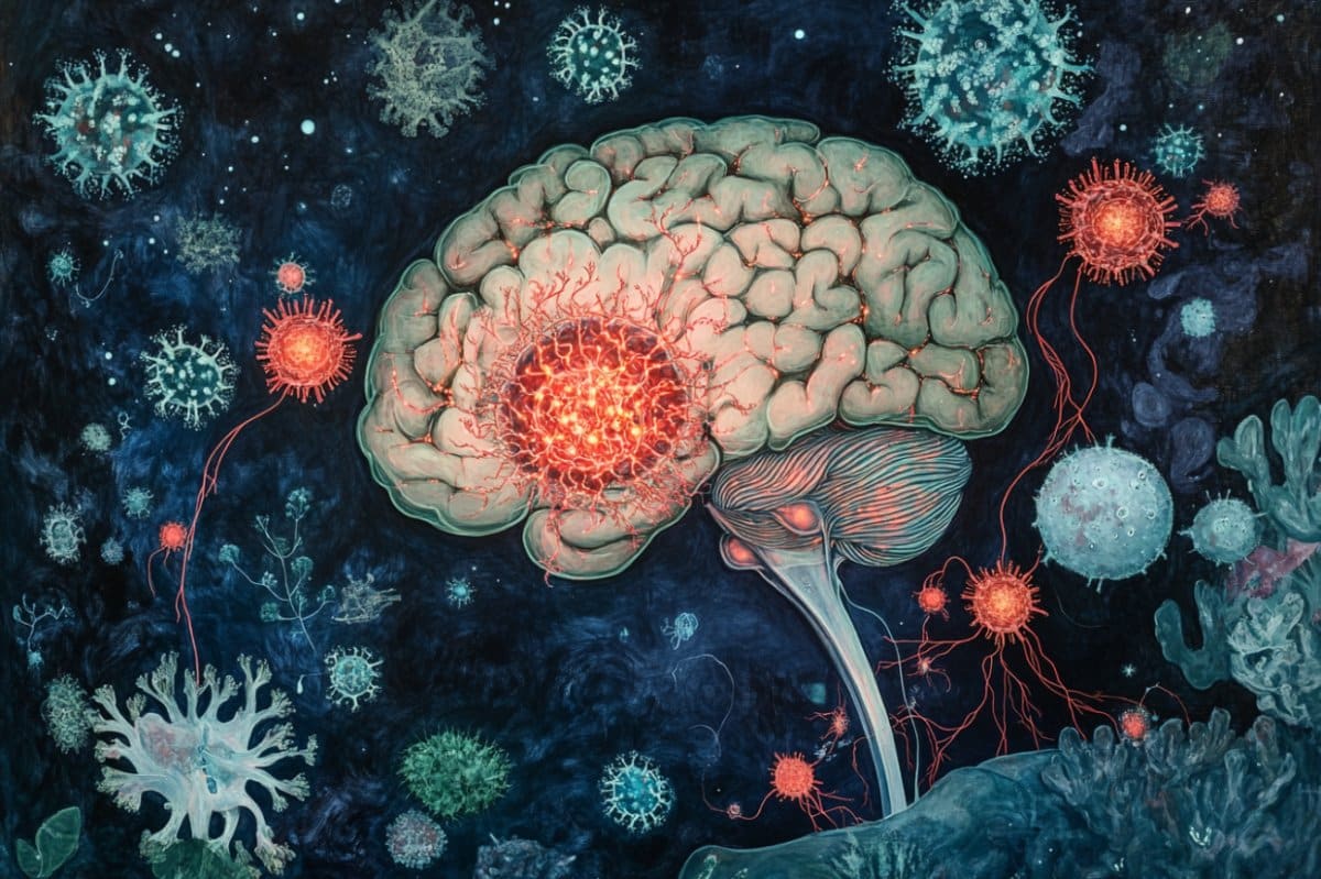
The brain is a unique place. It is protected by a large part of the body by the blood-brain barrier, which means that it is protected by pathogens and potentially dangerous substances that could be in our blood.
And historically, many scientists also believed that separation extended to the immune system: the brain has its own specialized immune cells called microglies, but the immune cells present in the rest of the body have long been considered to avoid the brain unless there is a disease or other problem requiring its presence.
Now, a team of scientists from the Yale School of Medicine (YSM) has shown that immune cells known as T cells reside in the healthy brain of mouse and humans, have come from intestine and fat. This is the first time that T cells have lived in the brain under normal and unprecedented conditions.
The results were published on May 28 Nature.
The presence of T cells in the healthy brain and proof that they travel between the brain and other parts of the body have turned the dogma of the field over the role of T cells in the brain, according to the authors of the study. Pathologists have already seen T cells in the brain, but it is assumed that they were there to respond to current or previous infections.
“We consider T cells as something fighting against infections and causes an autoimmune disease, but the surprise of this study is that T cells have a different role in the biology that we were not aware,” explains David Hafler, MD, William S. and Laws Stiles Professor of Neurology in YSM.
“With this article, we have definitely shown that their presence is not only linked to the disease but is part of normal physiology, and that it changes everything.”
The researchers found the most densely concentrated T cells in a small region called subngical organ, which is deeply nestled inside the brain and is known to regulate thirst and hunger.
The team found the cells in the subngical organs of laboratory mice and deceased and gave their brain to science.
This part of the brain also has a slightly fleeing blood brain barrier, which, according to researchers, allows cells in this region of the brain to more easily receive blood signals to find out when its host animal has to drink or eat.
“This makes it all the more interesting as the immune cells are also there, probably to provide a kind of signal on the normal state of the body,” explains Tomomi Yoshida, doctoral student YSM and the first author of the study. Yoshida managed the work with Andrew Wang, MD, PHD, Associate Professor of Internal Medicine and Immunobiology and Hafler.
Relay intestine signals to the brain
T cells fulfill many different functions, and researchers often classify this wide class of more-ended immune cells depending on the types of protein they display on their surfaces.
Looking at the brain’s T cells through this lens, YSM researchers found that they were different from the T cells present in the membrane surrounding the brain and were most similar to those that reside in the intestine and adipose tissues.
In mice, the modification of the intestinal microbiome assigned the transport of these T cells to the brain. The researchers noted that when babies mice seed and started solid foods, the corresponding gap in their intestinal microbiobiles launched the TV from the intestine to the brain.
The mouses raised in an environment without germ, without intestinal microbiomas, had no T cells in their brain. And exhausting animals from brain T cells has changed their food search behavior when the mice was offered food after a quick short.
Researchers believe that immune cells can point out body status to the brain through a form previously unknown to intestinal brain communication.
The information route between the digestive system and the brain, also called axis of the intestine, plays many important roles in our health and our well-being, but previous studies had not discovered a direct route so that immune cells enter the brain of the intestine.
The other intestinal means of communication include the vagus nerve, which directly connects the brain to the intestines, and small molecules secreted in the blood. Based on the dissemination of molecules in the blood has struck Wang as an ineffective communication method.
“According to a principle of design, it seems a kind of risky means of sending information,” he said.
“How cool if you really reprogrammer an immune cell to represent the state of the intestine and then send this cell to the brain, where it can tell the brain what it has seen and allow the brain to adapt according to this information?”
This is exactly what Wang and his colleagues think of happening. T cells could speak to the brain of the nutritional state of the body, but they could also transmit other important signals such as the state of the intestinal microbiome, the researchers have hypothesized.
Wang thinks they could stop in fat tissues on their intestine path to the brain as a kind of quality control point, but that remains to be tested.
Scientists then want to see how T cells know how to go from intestine to brain as well as what happens to these cells in neurological diseases such as multiple sclerosis or Parkinson’s disease.
“This study raises more questions than it answers,” says Yoshida. “But these are all interesting questions.”
Funding: The research reported in this press article was supported by the National Institutes of Health (Rewards R01AI162645, R01AR080104, P01AI073748, R01AI22220, UM1HG009390, P01AI039671, P50CA121974, R01CA227473 1F31NS130957-01A1, DP1DA050986 and R37AR40072) and the University of Yale.
The content is only the responsibility of the authors and does not necessarily represent the official opinions of the National Institutes of Health.
The research was also supported by the Smith Family Foundation, the Colton Center for Automunnity by Yale, The Food and Allergy Science Initiative, The Pew Charitable Trusts, The Mathers Family Foundation, The Ludwig Family Foundation, The Knights of Columbus, Race to Erase Ms, The Chan Zucerberg Initiative and The National MS Society.
About these news of research in neuroscience
Author: Rachel Timpa
Source: Yale
Contact: Rachel Timpa – Yale
Picture: The image is credited with Neuroscience News
Original search: Closed access.
“”The subngical organ is a nucleus for T cells derived from the intestine which regulates behaviorBy David Hafler et al. Nature
Abstract
The subngical organ is a nucleus for T cells derived from the intestine which regulates behavior
Specialized immune cells that reside in tissues orchestrate various biological functions by communicating with parenchymal cells.
The contribution of the innate immune compartment in the meninges and the central nervous system (SNC) is well characterized; However, the question of whether the cells of the adaptive immune system reside in the brain and are involved in maintaining homeostasis is not clear.
Here, we show that the subordinate organ (SFO) of the brain is a nucleus for T αβ parenchymal cells in the brain in the state of equilibrium in mice and humans.
Using the impartial transcriptomic, we show that these extravascular T cells in the brain are distinct from the t meninger cells: they secrete robust ifnγ and express tissue residence proteins such as CXCR6, which are necessary for their retention in the brain and for normal adaptive behavior.
These T cells are started in the periphery by the microbiome and the traffic of white and gastrointestinal adipose tissues towards the brain.
Once established, their number can be modulated by changes in the intestinal microbiota or the composition of the adipose tissue.
In summary, we note that CD4 T cells reside in the brain in the stationary state and are concentrated anatomically in the SFO in mice and humans; that they are transcriptionally and functionally distinct from the t meninger cells; And that they secrete ifnγ to maintain homéostasis of the SNC by omeostatic and intestinal homeostatic axes.
