A new study published in The Journal of Neuroscience Offers an overview of how small grooves on the surface of the brain – known as tertiary sulci – could help explain individual differences in reasoning capacity. Researchers from the University of California in Berkeley have found that the depth of these tiny cerebral folds in children and adolescents was linked to a stronger communication between two known areas to support higher order: the lateral prefrontal cortex and the lateral parietal cortex. The results suggest that subtle anatomical variations in these cerebral folds could influence how much different parts of the brain are during a complex thought.
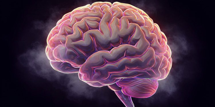
The outer layer of the brain, called cerebral cortex, is characterized by a structure folded with crests (gyri) and grooves (Sulci). These folds allow the large area of the cortex to register within the limits of the skull. While most research on neurosciences have focused on major sulci that are present in almost everyone, the present study focused on smaller and less deep grooves called tertiary sulci. These Sulci tend to vary more individuals and are found in areas of brain association – regions that have developed considerably during human evolution and are involved in advanced cognitive processes.
The justification of the study came from previous results showing that some Sulci in the lateral prefrontal cortex were associated with better reasoning skills in children and adolescents. By relying on this, the researchers have hypothesized that these small grooves could support reasoning by affecting the structure and function of the brain networks. More specifically, they proposed that deeper tertiary sulcis could bring the distant brain regions closer, allowing more effective communication – a concept known as the centrality of the network.
“I am interested in the way in which the prefrontal cortex, almost the third party before the human brain, supports cognitive functions of higher level such as reasoning, in coordination with its faithful parietal cortex and other regions,” said the author of the study Silvia Bunch, professor of psychology and member of the UC Berkeley’s Helen Wills Institute.
“I previously studied how the variation between individuals can be partially explained by the differences between them in terms of particular characteristics of anatomy and brain function. More recently, I have collaborated closely on a certain number of articles with my colleague Kevin Weiner, a neuroanatomist specializing in the Sulcal reference. ”
“Here, we wanted to rely on these results by testing whether the relationships we found between the sulcal depth and the performance of reasoning could be due to the fact that the Sulcal depth is substantial for the activity of the brain network.”
To examine this, the researchers recruited 43 neurotypical and adolescent children between 7 and 18 years of a broader development study of reasoning capacity. All the participants were native English speakers and underwent detailed brain imaging while performing a designed task to measure abstract reasoning. The task forced them to examine the models between simple forms and to determine relationships, both at a basic level (for example, by comparing forms) and at a more complex level (for example, identifying coherent rules between pairs of forms). The researchers also collected structural MRI analyzes to precisely map the Sulci in the brain of each participant.
A total of 42 sulci have been manually identified in each hemisphere of the brain, focusing on the prefrontal and lateral parietal cortex. The researchers then used functional MRI data collected during the reasoning task to assess the strength of each sulcus linked to others, essentially building a functional connectivity card through these grooves. Data analysis advanced techniques have been used to measure three aspects of the role of each sulcus in the cerebral network: the number of other sulcis to which it was connected (degree), how much it has served as a bridge between other (intermediate) connections, and how its connections have extended to different functional clusters (participation coefficient).
A key question was whether Sulci had unique connectivity models that distinguished them from each other. The answer was yes. Using automatic learning techniques, the team has shown that each sulcus had a very distinctive “digital connectivity imprint, which allows 96% accuracy to distinguish a sulcus from another according to its functional connections model. This strong discriminability argued that the Sulci are not only random anatomical wrinkles but can correspond to functionally significant brain units.
Then, the researchers brought together Sulci according to the similarity of their connectivity models. They found five major clusters, certain groups containing both prefrontal and parietal sulci and other compounds only with prefrontal sulci previously linked to reasoning.
Interestingly, some of these clusters were distinct from the large -scale brain networks generally identified in the studies of brain activity at rest. This suggests that using Sulci as a basic analysis unit could offer a more personalized and anatomically founded approach to study the brain function.
A key conclusion was that in several specific tertiary sulci – including the right PMFS -A, the left PMFS -I and the left PIMFS – a greater depth was linked to a higher network centrality. In other words, these deeper sulci tended to have stronger and more distributed connections on the brain network, in particular with other important sulci involved in vision and attention.
These relations were maintained even after taking into account the age and movement of the head, and were not motivated only by the proximity of the other Sulci. The results support the idea that a deeper tertiary sulci can promote greater neuronal efficiency by facilitating shorter and more direct communication between key brain areas.
“For some specific sulci, the more the sulcus is deep, the more it was integrated into the network of prefrontal and parietal sulci that we examined-that is to say, the more its activation was closely coupled with a number of other sulci.”
These associations were not uniform in all Sulci. Some sulci with large areas, such as those of intrapaietal sulcus, had in all higher centrality measures, but have not shown the same variation dependent on depth as tertiary sulci. The researchers also found that the link between sulcal depth and network connectivity was not confined to sulcites previously linked to reasoning capacity. For example, similar relationships have been observed in a sulcus called AIPSJ, which is sometimes used as a neurosurgical corridor, and a newly identified sulcus in the parietal lobe known as Slocs-V.
These results support a long -standing hypothesis that the individual variability of sulcal anatomy could play a functional role in cognition. The idea dates back to the 1960s, when neuroanatomistic sanids proposed only subtle differences in the sulcal structure could reflect and even shape cognitive capacities. Current results suggest that sulcal depth, especially in regions that develop at the end of gestation and continue to change during childhood, could serve as a biomarker for individual differences in reasoning and possibly other mental functions.
“Sulcal models are not random and are linked to cognition and functional organization. An individual fine-grained anatomy can help develop tools and found hypotheses to better take into account the high individual variability between people, “said a co-author Suvi Häkkinen, an assistant scientific project.
“We entered the study with the hypothesis that functional connectivity (that is to say the coordinated brain activation models through a set of regions) could be the missing link explaining the relationships between sulcal depth and cognition that we had already found,” added Bunge, but it was a nice surprise that turned out to be the relationship between depth and functional connectivity.
“I was surprised to learn from SUVI analyzes how a classifier of patterns could distinguish the sulci – even the small neighboring sulci – each other according to their fingerprints of connectivity (that is to say their coupling models to other regions).
Despite its strength, the study has several limits. The quantity of functional MRI data collected by participant was relatively modest, which can limit the accuracy of network measures at the individual level. The sample has also been limited to children and adolescents, leaving the question of whether similar models exist in adults or people with neurodevelopmental disorders. In addition, while the study used rigorous control analyzes to deal with confusion such as head movement and spatial proximity, certain residual effects cannot be excluded.
Future research could expand this approach to the different regions of the brain, age groups and cognitive areas. The method of definition of brain networks based on sulcal anatomy – rather than predefined regions or general brain atlas – can offer a more individualized means of studying brain function. It could also help identify early anatomical markers for cognitive forces and vulnerabilities, potentially illuminate education, clinical interventions and diagnoses based on neuroscience.
The study, “The anchoring of functional connectivity to individual sulcal morphology gives information in a pediatric study of reasoning“, Was written by Suvi Häkkinen, Willa I. Voorhies, Ethan H. Willbrand, Yi-Heng Tsai, Thomas Gagnant, Jewelia K. Yao, Kevin S. Weiner and Silvia A. Bunge.


