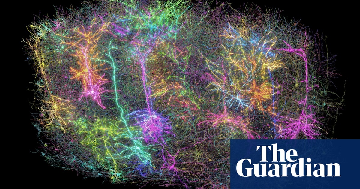
The most complete circuit diagram of neurons in a mammalian brain was created by scientists, providing revolutionary information on the mystery of the brain.
The map is of a visual cortex grain of a mouse, smaller than a grain of sand, and traces the structure of 84,000 neurons linked by half a billion synapses and approximately 5.4 km of neural wiring. 3D reconstruction of the cubic millimeter of the brain helps to discover how the brain is organized and how different types of cells work together, and could have implications for understanding intelligence, consciousness and neural conditions such as Alzheimer’s, Parkinson, Autism and schizophrenia.
Advanceds are “a moment of watersheds for neuroscience, comparable to the project of the human genome in their potential transformer”, according to Dr. David Markowitz, former program director of the American government organization Intelligence Advanced Research Projects Activity (IARPA), which coordinated the work.
The Microns project has not only sought to map the structure of neurons, but also studied electrical signaling between then, showing how they communicate and providing a better image of hidden conversations in the brain.
Scientists from the Baylor College of Medicine from Texas began by using specialized microscopes to record brain activity in the target region while the animal was watching various films and YouTube clips. Then, the researchers from the Allen Institute took this same cube millimeter from the brain and cut it in more than 25,000 layers, each 1/400th the width of human hair, and used a range of electron microscopes to take high resolution photos of each slice.
Finally, another team from Princeton University used artificial intelligence and automatic learning to rebuild cells and connections in a 3D volume. Combined, the massive data set is 1.6 petacts of size, equivalent to 22 years of non-stop HD video.
“Inside this little Speck is an entire architecture as an exquisite forest,” said Dr. Clay Reid, principal investigator and neurobiologist at the Allen Institute. “There are all kinds of connection rules that we know in various parts of neuroscience, and in the reconstruction itself, we can test the old theories and hope to find new things that no one has ever seen before.”
After promoting the newsletter
The results reveal new types of cells and a new principle of inhibition in the brain. Scientists previously thought of inhibitory cells – those that remove neural activity – as a simple force that reduces the action of other cells. But the latest works have revealed that inhibitory cells are very selective on the cells they target, creating a system of coordination and cooperation on the network scale.
Understanding the form and the function of the brain could open the way to a better understanding of the brain disorders involving disturbances in neural communication.
“If you have a broken radio and you have the circuit diagram, you will be better positioned to repair it.” said Dr. Nuno Da Costa, an investigator associated with the Allen Institute. “We describe a kind of Google card or plan of this sand grain. In the future, we can use it to compare cerebral wiring in a healthy mouse with brain wiring in a model of illness.”
The results are published in a series of articles in the journal Nature.
