There is an ongoing technological revolution that makes scientists faster and easier to see molecules that undermine human health – and possibly fight the problem.
The “resolution revolution” involves cryo-electron microscopes, whose still improved detectors and software produce three-dimensional images in unprecedented details, helping drug manufacturers.
They reveal so precise details that the biologist Andrew Ward, from Scripps Research to La Jolla, California, was able to highlight the Achilles heel of several types of coronavirus in the images he produced in 2016-2017.
The weaknesses he called? Spike Proteins – The now familiar elements that allow these viruses to infect cells.
This helped the manufacturers of medication, namely exactly what targets when Sars-Cov-2-The coronavirus that causes COVID-19-emerged at the end of 2019.
Ward produced an even clearer snapshot in proteins in 2020, helping scientists more to create vaccines.
With additional help from him, effective vaccines were quickly produced.
“It was just the start,” said Ward. “Now this technology opens doors that help us understand the roots of diseases such as cancer and neurodegeneration, including Alzheimer’s disease.”
“It regularly allows us to see the smallest machines in life – proteins, viruses and atoms they combine – with breathtaking clarity,” he added.
Research is not widely known to the public, in part because it is difficult to design how a microscope – not to mention that 10 feet high – can make a molecules’ moving gel, exposing their structure and their objective.
Ward offered a simple analogy to explain the question.
“Imagine entering a dark room,” he said. “You can almost say where the furniture is and see the dark contours of a sofa or a table. But once the lights are lit, you can view the color, texture, size and fine details. ”
This is what cryo -electric microscopes do – and with high speed.
Ward could only produce 200 images per day when he won his doctorate in scripps from 2003 to 2008, while he used a much less powerful cryo-electronic microscope type. And there was only access to the day or two every month.
Today, it can generate 1,500 images per second on Titan Krios, the largest and most powerful of the seven cryo-electron microscopes of scripps.
If you could stack the images he takes during a six-hour period, they would get up as high as Mount Everest, said Ward, who collaborated with institutes on Sars-Cov-2, Lassa, VIH, malaria and H5N1 bird flu.
With the unknown eye, the images seem weird. Some resemble a bumpy and frozen lava, other crumpled Christmas crowns. Still others look like knotty cords on old fixed phones.
But their importance is understood by scientists, especially those focused on preparing the world to the pandemic which could then come.
It is a bit of luck that Ward is a rising star in an institute that has helped develop more than 15 drugs and treatments approved by the FDA, including Humira, which is used by people with arthritis.
He was interested in science outside of Boston – less when he entered Duke University in the first year.
Things quickly changed when he took a job of work in a campus laboratory, where cellular biologists Michael and Mary Reedy have let tinn. Before a long time, Ward helped build the components of the cameras and microscope detectors, and was dazzled by what they could do.
“I started to see molecules and atoms,” Ward said 46 years old. “It sort of blows you to follow things to this resolution.”
Electron microscopes have existed since the 1930s, and they have played a vital role in the revelation of the protein and virus structure and how they work. But the instruments started to enter their current golden age only about 2001, the year when the district arrived at Scripps as a laboratory technician.
The advances have mainly reached the past decade, strongly improving the resolution of the image and allowing scientists to see individual atoms. Software has also facilitated molecules to interact with potential drugs, helping to determine which should go to large -scale clinical trials.
Boom, locally and in the world, has not gone unnoticed. In 2017, three European scientists won the Nobel Prize in chemistry for helping to transform cryo-electrons microscopes into essential tools to explore the life sciences.
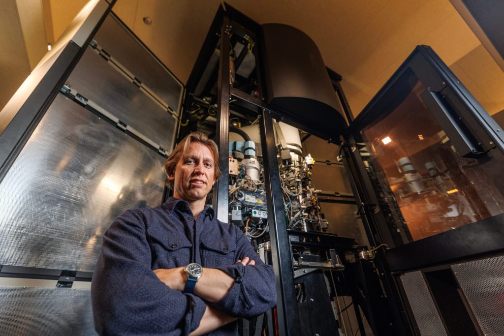
Ward says he is happy to be in the background. But he has become a leader in the field – mainly by using Titan Krios.
“Big Daddy”, as Ward calls it, is very sensitive. The imposing microscope is seated on stabilizers to prevent something as simple as a door slammed to produce vibrations that could spoil the image taking. It works in silence for the same reason.

In clear terms, the microscope freezes organic samples, then strikes them with bundles of electrons that create images.
“Once you see the layout of atoms, the connectivity of molecules, you can become an engineer,” said Ward. “You can move things and manipulate the constituent elements of life to make new therapies and vaccines that have a much higher probability of success compared to engineering without plans.
“We have accelerated the choice process of the one that should be a blow, or not, for clinical trials,” said Ward.
This does not mean that scientists are about to flood the market with new prevention means.
“Potential vaccines will have to go collectively from five to seven years of trials in humans,” he added. “But we no longer shoot in darkness or no longer rely on empiricism.
“We can now shine a light – or rather a very powerful bundle of electrons – on research on vaccines in scientific driving.”
California Daily Newspapers
