Scientists developed embryo-like structures in the laboratory that produced human blood cells, opening up new possibilities for regenerative medicine.
The ability to generate blood stem cells in the laboratory may one day make it possible to treat patients in need of bone marrow transplants using their own cells.
The advance is the latest in a rapidly evolving field in which embryo models are created from stem cells without the need for eggs or sperm, opening a window into the earliest stages of human development.
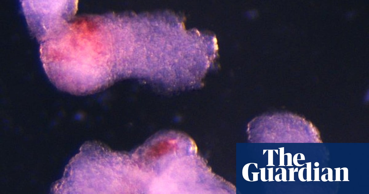
“It was an exciting moment when the blood-red color appeared in the dish – it was visible even to the naked eye,” said Dr Jitesh Neupane, a researcher at the Gurdon Institute at the University of Cambridge and first author of the study.
He and his colleagues use the model system to understand the early stages of heart and blood development.
“This sheds light on how blood cells form naturally during human embryogenesis, offering potential medical advances for testing drugs, studying early blood and immune development, and modeling blood disorders like leukemia,” Neupane said.
Human stem cells used to develop embryonic structures can be created from any cell in the body. This means that this approach could also pave the way for producing blood that is fully compatible with the patient’s body.
Although there are other methods for generating human blood stem cells in the laboratory, these require a cocktail of additional proteins, while the new method mimics the natural developmental process in which self-organized structures lead to the formation of different cell types.
“Although still in its early stages, the ability to produce human blood cells in the laboratory marks an important step towards future regenerative therapies – which use a patient’s own cells to repair and regenerate damaged tissues,” said Professor Azim Surani of the Gurdon Institute, lead author of the paper.
In this latest study, scientists used human stem cells to replicate certain cells and structures that typically appear during the third and fourth weeks of pregnancy. The model was specifically designed to not contain the tissues that will form the placenta and yolk sac in a natural embryo, meaning it did not have the theoretical potential to develop into a fetus and did not develop the tissues that would form the brain.
“It’s a minimalist system,” Neupane said.
The team observed the emergence of three-dimensional structures resembling embryos under a microscope. By the second day, they had self-organized into three germ layers – called ectoderm, mesoderm and endoderm – which constitute the foundations of the human body plan. By the eighth day, beating heart cells had formed, cells that would eventually give rise to the heart of a developing human embryo.
On day 13, the team saw red spots of blood appear. Blood stem cells taken from the model were also shown to be able to differentiate into different types of blood cells, including oxygen-carrying red blood cells and white blood cells, which are essential for the immune system.
The results are published in the journal Cell Reports.